Annual comprehensive eye examinations are a vital part of preventive eye health care. Adults and children alike are often unaware there are any existing problems since there are no obvious signs or symptoms in many ocular issues. Comprehensive eye exams are extremely important for creating a baseline, maintaining good vision and eye health, and to help prevent potential vision loss with early diagnosis and treatment of various eye conditions.
Call 469.490.ENVY (3689) today or schedule an appointment online for your comprehensive eye exam.
Early detection is key to help prevent vision loss. Since many vision issues show no obvious signs or symptoms, the only way to truly get an overall health check of your eyes is by seeing a doctor of optometry for a check-up.
The doctors at Envy Eye Care will also go over your overall health and family history, eye or vision symptoms, medications, and potential work-related or environmental issues that may affect your vision. You will also have a preliminary test such as depth perception, color vision, eye muscle movements, peripheral or side vision, and how your pupils respond to light.
Getting a comprehensive eye exam which includes dilation drops or retinal imaging is the only way to visualize the blood vessels behind the eye for issues without an x-ray, CT scan or invasive surgery.
The doctors at Envy Eye Care use a wide variety of tests and procedures to examine your eyes. These tests range from simple ones, like having you read an eye chart, to complex tests, such as using a high-powered lens to visualize the tiny structures inside of your eyes.
When you get a comprehensive eye exam at Envy Eye Care, these are the equipment that your optometric technician and doctor will use when checking your eye health.
Non-Contact Tonometer or iCare Tonometer
Many know the tonometer machine as the "air puff machine" because it blows a little puff of air into your eye. This machine checks the pressure of your eyeball and can help diagnose glaucoma.
iCare tonometer is an instrument that uses rebound technology that “tickles” the front of your eye so you barely feel it. It's an alternative for the patient who is timid of the air puff.
Auto-refractor
Auto-refractor keratometer will create a baseline for your prescription.
Visual Field Test
Visual field test is a method of measuring an individual's entire scope of vision, that is their central and peripheral vision.
Optomap
A non-dilating camera that captures up to a 200 degree digital image of the retina.
Digital Eye Exam with CV5000
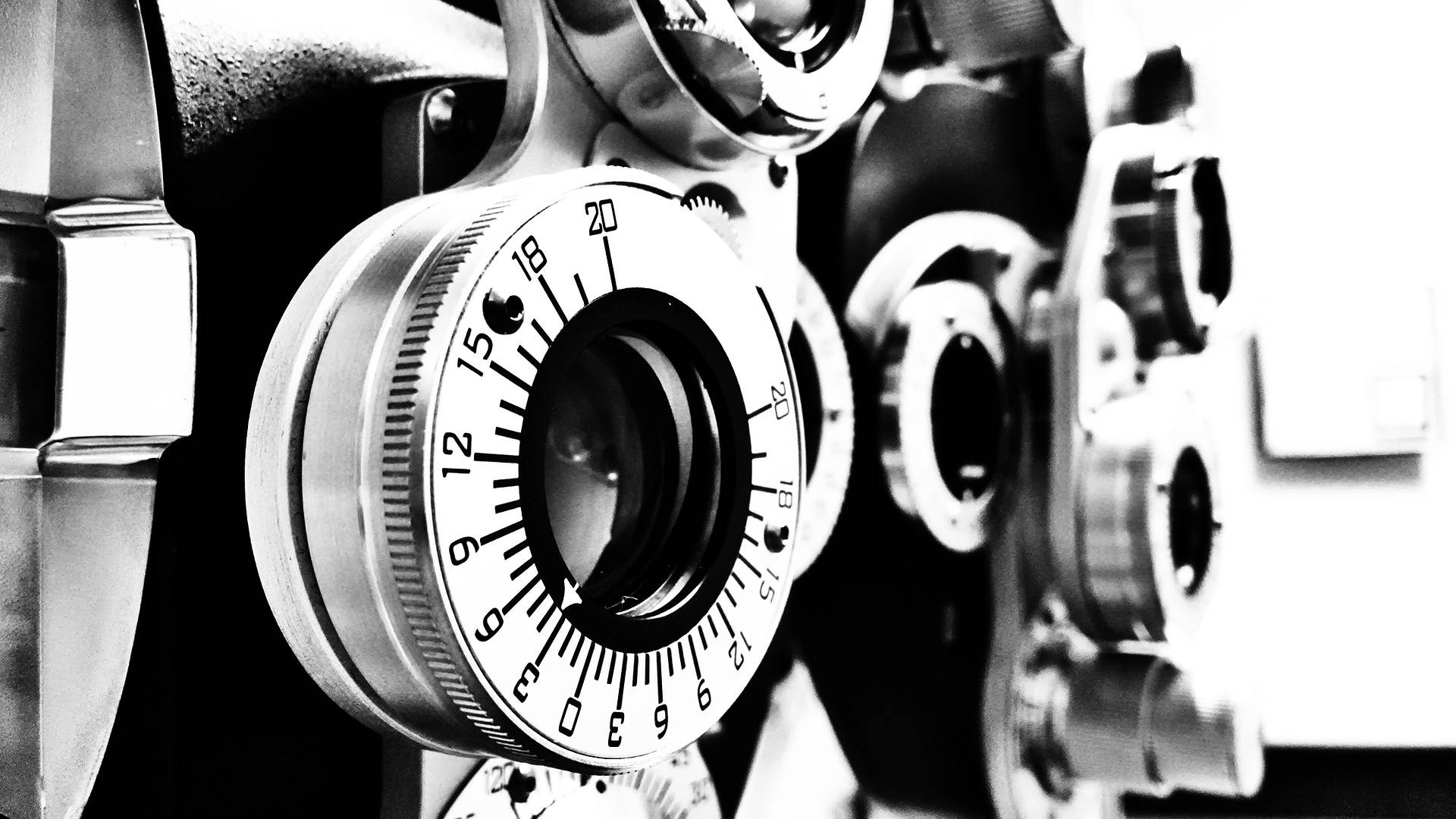
The CV5000 automated phoropter offers all the latest features in automated refraction.
Slit Lamp
A slit lamp is a binocular microscope that your doctor uses to examine the structures of your eye under high magnification. A wide range of eye conditions and diseases can be detected with the slit lamp exam.
Keratometer
This machine measures your cornea's shape. If your cornea is more round or flatter, this will determine whether or not you have astigmatism or corneal problems. For contact lens wearers, this machine assesses the shape of the eye to find the proper contact lens fit.
Optical Coherence Tomography (OCT)
OCT is often used to evaluate disorders of the optic nerve and/or retina. The OCT exam helps your doctor see changes to the fibers of the optic nerve. OCT is a non-invasive imaging test and it uses light waves to take cross-section images of your retina.
Corneal Topography
Corneal topography is a computer assisted diagnostic tool that creates a 3-D map of the cornea's shape and curvature. It enables detection of corneal diseases, and irregular corneal conditions, such as swelling, scarring, abrasions, deformities, and irregular astigmatisms. This type of analysis provides your doctor with very fine details regarding the condition of the corneal surface. These details are used to diagnose, monitor, and treat various eye conditions.
Pupil dilation has been the traditional standard of care. For most people, retina imaging isn't required. However, it does provide another tool for assessing retina and eye health, which can be helpful during technically difficult examinations.
Dilation Drops
• Drops are instilled into the eyes
• Takes 20-30 minutes for the dilations to take effect before the doctor could continue with the exam
• Sides effect includes blurry vision up close and light sensitivity up to 6 hours
Optomap® Retinal Imaging
• A quick and easy scan that captures a digital image of the retina
• An enlarged image to see more detailed view of the retina
• Immediately available to view and discuss during your exam
• A permanent record for your medical file which allows for future comparisons
• A good alternative for those who cannot or do not want the dilation drops
• There are no sides effect such as light sensitivity and blurry vision
Whether eye dilation during an exam is necessary depends on the reason for your exam, your age, your ethnic background, your overall health and your risk of eye diseases.
A thorough, dilated exam allows the doctor to do a complete exam of the retina, and that is important to do throughout your life, as several eye diseases and conditions are detected at their earliest stages during a thorough eye exam.
Common eye diseases that may be identified and treated include:
• Diabetes
• High Blood Pressure
• Macular Degeneration
• Retinal detachment
• Glaucoma
Common systemic conditions like high blood pressure, diabetes, and high cholesterol can be recognized and referred to the appropriate specialist.
AI Website Software